By Vidja Rajan, Columnist, The Times
The purpose of circulation is to bring nutrients to cells and remove wastes. Circulation is important for all organisms, including plants, fungi and bacteria; it’s no good to a cell if materials cannot get in and out efficiently. In single-celled organisms, which live suspended in an aqueous environment, the medium performs this function.
For single-celled organisms that grow on solid surfaces such as bacteria in your mouth, only the top layer is supplied with nutrients, and the lower layers consequently die. Thus, plaque builds up layer upon layer, especially in areas such as between and behind teeth that are not cleaned regularly. Over time, plaque can develop a crusty mineralized layer called tartar, cementing the dead bacteria to the substrate, with living layers on the surface of the biofilm where saliva circulates materials. By contrast, rather than having a superficial or external circulatory system, multicellular organisms, which contain many layers of cells, enclose the circulatory system within the body such that all layers are serviced. In animals, circulatory systems come in two forms: open and closed.
An open circulatory system washes the surface of each cell of the body. In vertebrates, this open system is the lymphatic system and in invertebrates it is the hemocoel. Because phagocytic immune cells circulate in the open circulatory systems, this process allows the policing of the interstitial space between cells that may get contaminated by injury. In vertebrates, lymph drains into the lymphatic system and gets filtered through lymph nodes. The lymph nodes are where lymphocytes, immune cells of the T-cell and B-cell ilk, have a chance to interact with and distinguish between their own and foreign materials. Body cells that get washed into the lymph get a pass, but foreign bodies like bacteria and viruses that enter get trapped and destroyed. This is also the reason why many vaccinations are given as a shot in the arm – the vaccine materials can get efficiently directed by lymphatic currents into the lymph node, where they can prime B- and T-cells. This is also the reason why your tonsils and adenoids, the major lymphatic regions of the neck, swell up when you have an upper respiratory tract infection.
The open circulatory system is more ancient than the closed system, and exists in all animals, from evolutionarily ancient primitive sponges and jellyfish to recent vertebrates. In insects such as bees, interstitial space is washed by the hemocoel, which delivers nutrients and removes wastes. But transportation of oxygen is not a high priority for the hemocoel, since respiratory trachea deliver oxygen directly to cells. Nonetheless, some invertebrates also contain an oxygen-binding, metal-containing protein called hemocyanin. This metallo-protein is conjugated to copper, rather than iron as in hemoglobin. Rather than being carried on cells, hemocyanin is dissolved directly in the hemocoel. Other oxygen-binding pigments in invertebrates are hemerythrin and chlorocruocin. I will discuss these pigments a little more at the end of this article. The hemocoel also contains phagocytic immune cells called hemocytes, which engulf and destroy foreign bodies in a manner similar to the white blood cells of vertebrates.
The hemocoel in insects is analogous to a bag full of liquid sloshing about. The problem is that sloshing is not an effective mechanism for efficiently circulating liquid around the body. Circulation of hemolymph is improved by the action of muscle contraction and also by actively pumping it from the posterior to the anterior using a tube open on both ends, with a series of contractile “hearts” that move the liquid in the tube forward. Hemocoel enters the tube by being drawn in through openings in the hearts called ostia (see Figure 1). The fluid is then shunted towards the head where it oozes out and flows backwards to the rear of the animal. Interestingly, in the open circulation of some vertebrates, like fish, amphibians and some birds, lymph hearts perform the same function. In other vertebrates, such as mammals, muscle contraction helps to drive lymph movement. If you sit unmoving for a long period of time, such as on a flight, lymph pools in the feet, causing edema. Flexing your leg muscles helps with circulation of lymph in these situations.
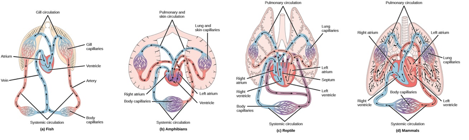
In vertebrates there is a second and more familiar system, the closed blood circulatory system. A few invertebrates, the annelid worms (earthworms) and the cephalopod molluscs, such as octopus, also have a closed system, but they are unusual in having this advanced one-way system. To understand this statement, consider that having clear directionality allows high efficiency. A closed circulatory system carries oxygenated blood to the tissues of the body through the arteries and removes the wastes through the veins. This one-way system is highly efficient, because it prevents the mixing of nutrient-rich and nutrient-poor blood. In vertebrates, oxygenation takes place in either gills/skin in aquatic organisms, or the lungs in terrestrial organisms. The number of pumps in the closed circulatory system varies from two to four. The heart is a folded tube which evolved in stages to push blood to the pulmonary regions where oxygenation takes place, and then around the body. Fish have a one-loop circulation which takes blood directly from the gills where it is oxygenated to the body where oxygen is delivered to body cells. Two-loop circulation evolved by degrees, and represents one pulmonary loop where blood is oxygenated, and one systemic loop where it is deoxygenated, with the heart as the nexus between the two. See Figure 2 for examples of the closed circulatory system of fish, amphibians, reptiles and mammals.
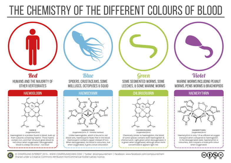
There are a plethora of pigments that carry oxygen around the body in a rainbow of colors: red, blue, green, and violet (see Figure 3) [1]. The vertebrate’s red pigment, hemoglobin, is a metallo-protein with an iron core, with the ability to carry four oxygen molecules at a time. Hemocyanin is blue and is a large molecule associated with copper dissolved directly in the hemocoel that binds and releases oxygen less well than hemoglobin. This is not such a big problem as it might appear on the surface, because insects also have tracheal tubes that go from spiracles to cells, carrying out gas-exchange functions. Still, many invertebrates such as octopuses have very high levels of hemocyanin just to move oxygen around. Some animals have green blood. there are two separate reasons for green blood: the presence of the pigment chlorocruorin in some annelids, and biliverdin found in the green-blooded skink. Biliverdin is a breakdown product of hemoglobin which colors the skink just as chlorophyll, a chemical cousin of hemoglobin which contains magnesium rather than iron, colors plants. Finally, there is violet hemerythrin, which is associated with free floating cells (corpuscles) in the hemocoel of some marine worms and a group of ancient invertebrates called bryozoans.
Finally, the chemical origins of the pigment hemocyanin is from the enzyme tyrosinase, an ancient enzyme that has a particular affinity for oxygen and is also part of the synthetic pathway for the pigment melanin. This implies that the origins of pigments are as a protective antioxidant mechanism during prehistoric times when the levels of oxygen in the atmosphere jumped massively, causing the “Great Oxygen Event”, a catastrophe for the anaerobic lifeforms, that occurred about 2.5 billion years ago. With these pigments to provide protection from oxygen, just as melanin provides protection from the sun, the organisms went on to leverage it to supply tissues with oxygen, aiding the evolution of more complex life forms, such as insects and us.
- Lutz, D., The Many Colors of Blood. Chem Matters, 2010.
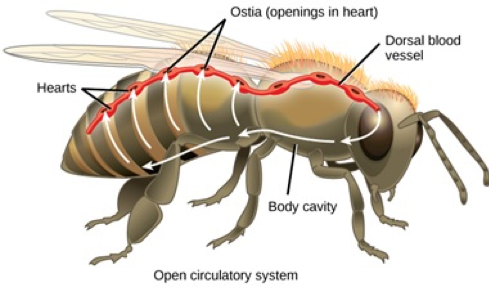
Figure 1: The Open Circulatory System of a Honeybee
Circulation in the hemocoel in invertebrates is assisted by the transport of liquid into the dorsal blood vessels through openings in the contractile hearts called ostia. The hemocoel seeps out of the front opening and flows to the posterior.
Credits: Figure from LibreTexts. Content is licensed by CC BY-NC-SA 3.0. https://bio.libretexts.org.
Figure 2: The Closed Circulatory System showing the gill/pulmonary circuit where blood is oxygenated and the systemic circuit where blood is deoxygenated. The sequence from left to right shows: a) fish with a single loop systemic circulation where blood leaves the gill and goes straight to the body, b) amphibians with two loops but with a heart where oxygenated and unoxygenated blood mix in both upper and lower chambers, c) reptiles with two circulations with oxygenated and unoxygenated blood mixing in only the upper chamber of the heart, d) mammals (and birds) with two completely separated loops carrying oxygenated and unoxygenated blood with no mixing in the heart. Modified from: https://bio.libretexts.org/ LibreTexts content is licensed by CC BY-NC-SA 3.0.

Figure 3: The many pigments in animal blood. Animal blood appears to have evolved as an antioxidant, and then was recruited to transport oxygen around the body. Blood also carries dissolved or suspended nutrients to the cells and removes metabolic wastes.
Credits: Reproduced under a Creative Commons Attribution License from https://www.compoundchem.com/2014/10/28/coloursofblood/